CASE20240716_009
[Invited Case] PPVI in Young Man S/p Repair of TOF with RVOT Aneurysm
By Supaporn Roymanee, Thanawat Suesat
Presenter
Thanawat Suesat
Authors
Supaporn Roymanee1, Thanawat Suesat2
Affiliation
Prince of Songkla University, Thailand1, Khon Kaen Hospital, Thailand2,
View Study Report
CASE20240716_009
Pulmonic Valve Intervention - Pulmonic Valve Intervention
[Invited Case] PPVI in Young Man S/p Repair of TOF with RVOT Aneurysm
Supaporn Roymanee1, Thanawat Suesat2
Prince of Songkla University, Thailand1, Khon Kaen Hospital, Thailand2,
Clinical Information
Relevant Clinical History and Physical Exam
19 year old male ,Known case TOF s/p total repair since 2007
- Heart: no heave, no thrill, normal S1, S2, to and fro at LUPSB


- Heart: no heave, no thrill, normal S1, S2, to and fro at LUPSB


Relevant Test Results Prior to Catheterization
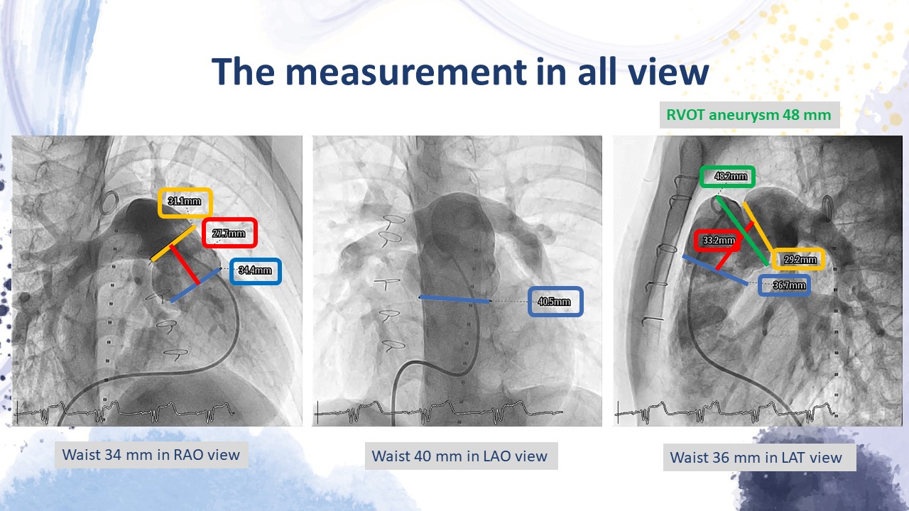
PA gram : Waist 34 mm in RAO view , Waist 40 mm in LAO view , Waist 36 mm in LAT view , RVOT aneurysm 48 mmThe right femoral vein is patent with a diameter of 13.5 mm
 PA1.mp4
 PA2.mp4
 PA3.mp4
Relevant Catheterization Findings
Interventional Management
Procedural Step
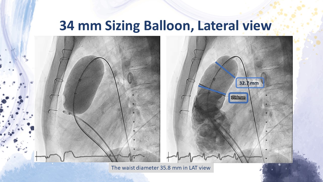
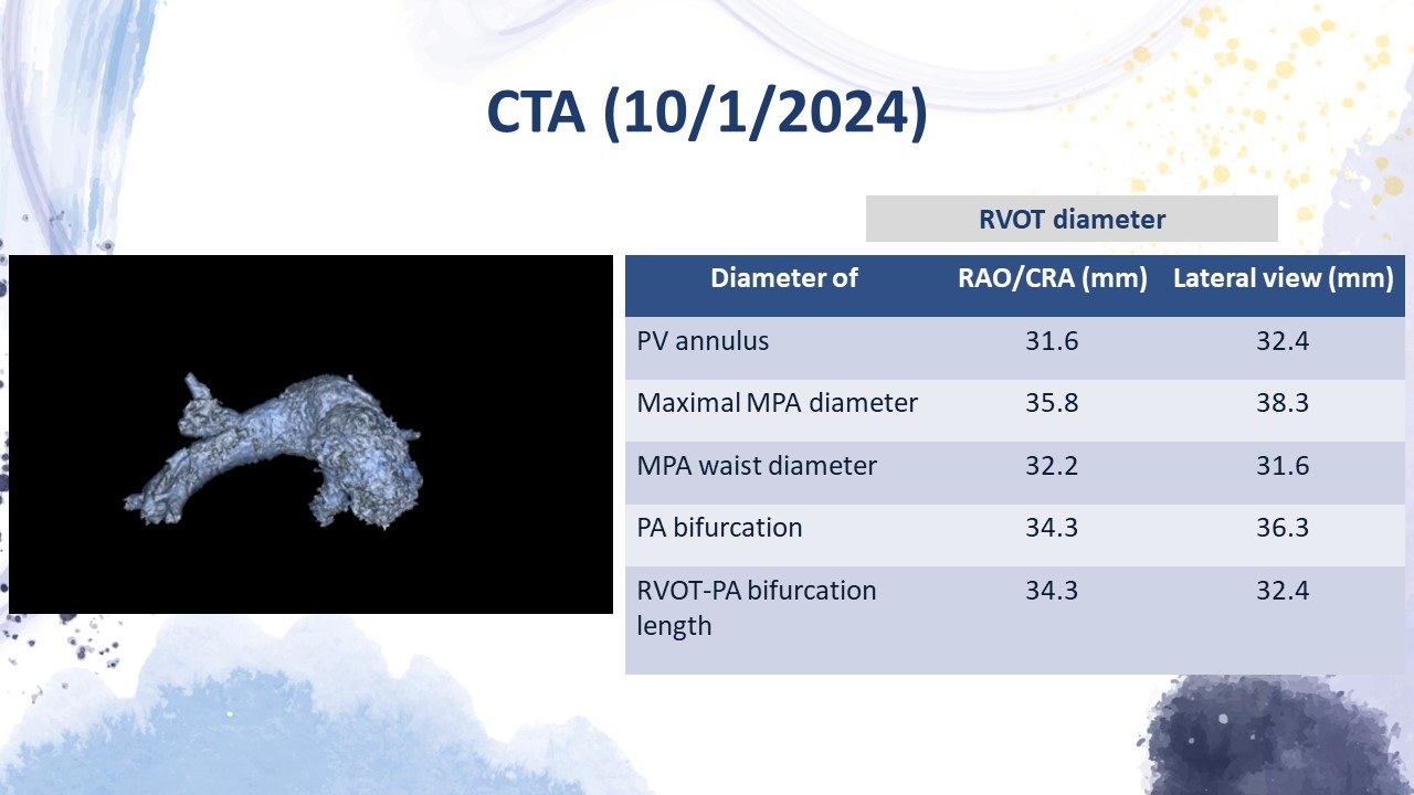
1. Preclosed Proglide was done before procedure2. MPAangiogram in RAO cranial and true lateral view show pyramidal shape RVOT withwaist dimeter 36x36 mm , bifurcation 29.5 x 29.5 mm , length 28.5 x 29.4 mm3. With 34mm balloon sizing , waist dimeter 29 x 31 mm , No coronary compression duringballoon interrogation4. 26 Frlong Gore DrySEAL was inseted to PA branch , Venus P valve 36/25 was selected to implant at RVOT5. After procedure , RV gram and PA angiogram show good device position , No PR , Nosignificant PA-RV gradient , No obstruction at both PA branch6. CAG show no coronary obstructionComplication : none
 8.2.mp4
 13.2.mp4
 16.2.mp4
Case Summary
Percutaneous pulmonic valve implantation using the newself-expandable Venus P-valve proved to be a safe and viable procedure,enabling the treatment of highly dilated right ventricular outflow tracts with aneurysmal change that are unsuitable forexisting balloon-expandable valves.
