CASE20240524_004
Exploring Abnormalities Along the Path, Capturing the Flying Calcium
By Ho On Alston Conrad Chiu, Cheung Chi Simon Lam, Tai-Leung Daniel Chan, Eric Kwong Yue Chan
Presenter
Ho On Alston Conrad Chiu
Authors
Ho On Alston Conrad Chiu1, Cheung Chi Simon Lam1, Tai-Leung Daniel Chan1, Eric Kwong Yue Chan1
Affiliation
Queen Mary Hospital, Hong Kong, China1,
View Study Report
CASE20240524_004
TAVR - Complex TAVR
Exploring Abnormalities Along the Path, Capturing the Flying Calcium
Ho On Alston Conrad Chiu1, Cheung Chi Simon Lam1, Tai-Leung Daniel Chan1, Eric Kwong Yue Chan1
Queen Mary Hospital, Hong Kong, China1,
Clinical Information
Relevant Clinical History and Physical Exam
Our patient is an 80-year-old man with history of lympohma, previously received chemotherapy and radiotherapy. He presented with dyspnea on exertion and recurrent syncope. Ejection systolic murmur was noted at the right upper sternal border with radiation to the neck. Referred for consideration of TAVR in view of echocardiographic findings of severe aortic stenosis (Aortic valve area of 0.8cm2 and Aortic valve gradient of 75/46mmHg).
 Severe AS combined.mp4
Relevant Test Results Prior to Catheterization
Subsequent CT analysis noted an abnormal sub-valvular lesion. To further evaluate the sub-valvular lesion, reassessment trans-thoracic echocardiography (TTE) and trans-esophageal echocardiography (TEE) - with 3D reconstruction - were performed. Reassessment echocardiography identified a densely calcified aortic valve with hypoattenuated signal at the left ventricular outflow tract level, with findings suggestive of a fibromusclar ridge. A highly mobile calcification was also identified on TEE.
Subvalvular Lesion Combined.mp4
Mobile Calcification Combined.mp4
Relevant Catheterization Findings
Cardiac catheterization was perfomed. Coronary angiography with JL4 and JR4 diagnostic catheters showed only minor coronary artery disease only. Aortogram via 5Fr pigtail catheter was also performed prior to our TAVR procedure for evaluation of aortic arch anatomy prior to our procedure. Best alignment angle was also determined, including 3-cusp and cusp overlap views.
coronary angiography.mp4
normal Aortogram.mov
Interventional Management
Procedural Step
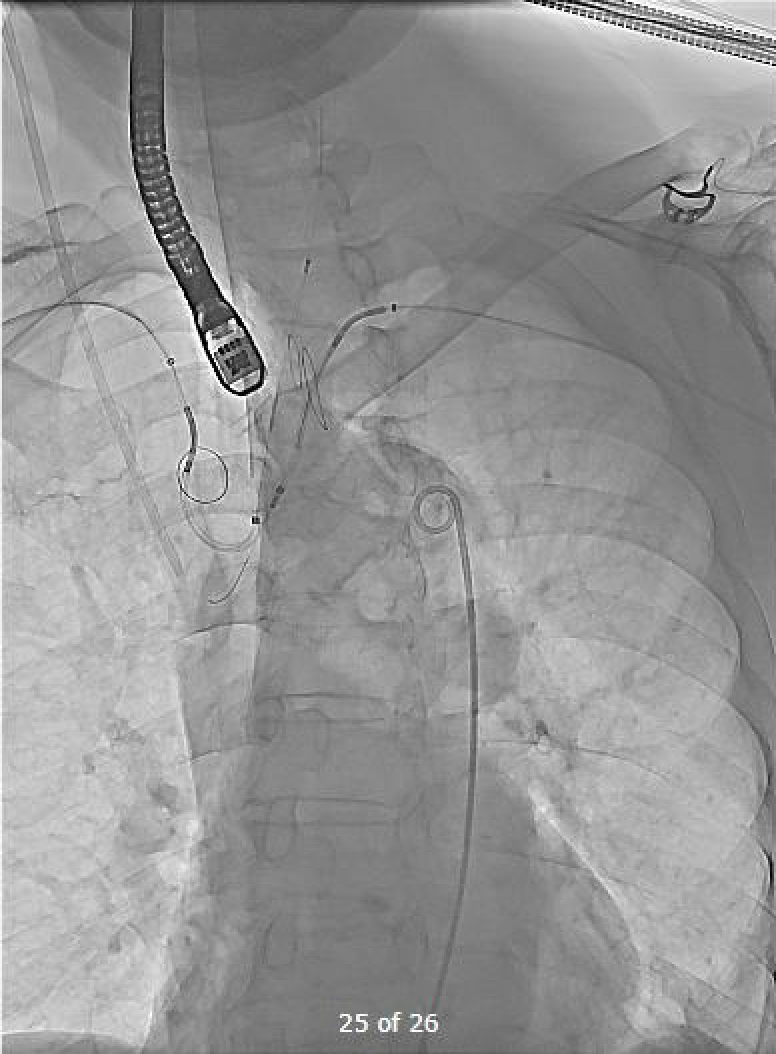
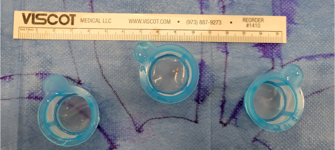
Heart team decision was to proceed with transfemoral TAVR (Evolut FX 34mm) with double Sentinel Cerebral Embolic Protection.Vascular accesses were established in the following fashion: (a) pigtail catheter insertion via a 6Fr sheath at left femoral artery, (b) temporary pacing line insertion via a 6Fr sheath through the left femoral vein, and (c) main access via the right femoral artery with final introduction of an 18Fr Sentrant Sheath.For our "Double Sentinel Cerebral Protection" strategy, the Sentinel devices were deployed in the following manner: (1) two filters were deployed at the brachiocephalic artery and left carotid artery via right radial access in standard fashion, (2) an additional filter was deployed at the left subclavian artery via left radial access.The aortic valve (AV) was crossed with a Straight Tip Emerald guidewire via an AL1 catheter. After crossing the AV, we exchanged to a Safari Small guidewire, with excellent wire position achieved. Pre-dilatation with TRUE Balloon 24mm x 4.5cm under rapid pacing was performed, and a TEE image demonstrating embolization of the highly-mobile calcified lesion was acquired. Evolut FX 34mm was deployed successfully. Further post-dilatation with TRUE Balloon 26mm x 4.5cm was done, valve frame expansion was then optimized. Final TEE and angiographic results were excellent, with the subvalvular lesion being pushed aside. Filters from the Sentinel devices also showed significant debris, particularly the distal RRA filter.
flying Calcium Combined.mp4
Post-dilatation Fluoro and TEE.mp4
Case Summary
This case demonstrated unusual but interesting findings along the path of left ventricular outflow tract, from the subvalvular to the valvular level. Pre-procedural imaging findings, from echocardiography (especially with 3D reconstruction) and computer tomography in our case, may help to identify patients who are at risks of cardioembolic events. Our "Double Sentinel Cerebral Protection" strategy may also be an adoptable strategy in preventing catastrophic cardioembolic events from taking place, particularly in patients with high-risks pre-procedural imaging findings as demonstrated in our case.