CASE20220613_001
A Pilot Challenging Study Of Early 3-Tesla Cardiac Magnetic Resonance Imaging Following TAVR Within 14 Days: Safety, Image Quality, And Long-term Clinical Outcomes
By Numfon Tweeatsani, Ammarut Chuajak, Adun Kampaengtip, Chonthicha Tanking, Wongsakorn Luangphiphat, Viroj Muangsillapasart, Komen Senngam, Anuruck Jeamanukoolkit, Tanyarat Aramsareewong, Jiraporn Laothamatas
Presenter
์Numfon Tweeatsani
Authors
Numfon Tweeatsani1, Ammarut Chuajak2, Adun Kampaengtip3, Chonthicha Tanking4, Wongsakorn Luangphiphat5, Viroj Muangsillapasart5, Komen Senngam5, Anuruck Jeamanukoolkit6, Tanyarat Aramsareewong7, Jiraporn Laothamatas1
Affiliation
Chulabhorn Royal Academy, Thailand1, Queen Savang Vadhana Memorial Hospital, Thailand2, AIMC ramathibodi hospital Mahidol university, Thailand3, Chulanhorn Royal Academy, Thailand4, Chulabhorn Hospital, Thailand5, Police General Hospital, Thailand6, Phramongkutklao Hospital, Thailand7
Valve - Others (Valve)
A Pilot Challenging Study Of Early 3-Tesla Cardiac Magnetic Resonance Imaging Following TAVR Within 14 Days: Safety, Image Quality, And Long-term Clinical Outcomes
Numfon Tweeatsani1, Ammarut Chuajak2, Adun Kampaengtip3, Chonthicha Tanking4, Wongsakorn Luangphiphat5, Viroj Muangsillapasart5, Komen Senngam5, Anuruck Jeamanukoolkit6, Tanyarat Aramsareewong7, Jiraporn Laothamatas1
Chulabhorn Royal Academy, Thailand1, Queen Savang Vadhana Memorial Hospital, Thailand2, AIMC ramathibodi hospital Mahidol university, Thailand3, Chulanhorn Royal Academy, Thailand4, Chulabhorn Hospital, Thailand5, Police General Hospital, Thailand6, Phramongkutklao Hospital, Thailand7
Clinical Information
Relevant Clinical History and Physical Exam
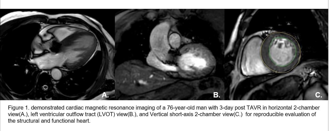
Cardiac magnetic resonance (CMR) imaging was widely and continuously used in imaging workflow for cardiac evaluation of both anatomical and functional issues over decades providing more information with reproducible measurement. No declarable optimal timeline and a lack of information on both safety and clinical efficacy for immediate CMR imaging following TAVR despite MRI safety being approved that all heart valve prostheses can undergo
MRI immediately after valve placement.
Our study aimed to assess safety, image quality, and long-term clinical outcome by using a 3-Tesla cardiac MRI following TAVR within 14 days in patients diagnosed with severe symptomatic aortic stenosis(AS).
Relevant Test Results Prior to Catheterization
Relevant Catheterization Findings
Interventional Management
Procedural Step
METHOD
 movie-0001.avi
 movie-0002.avi
 movie-0003.avi
 movie-0004.avi
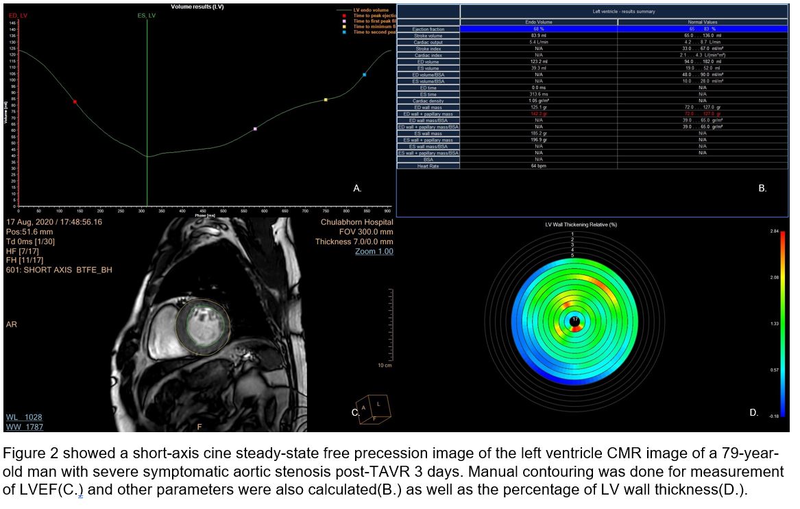
Cardiac MRI was performed using Philips 3T MRI machine at Diagnostic Imaging and Image-guided Therapy Centre for The People(DITP), Chulabhorn Chalermparkiet Medical Center, Chulabhorn hospital from November 2019 to December 2020 following MRI safety guidelines with ethical approval. Simultaneous monitoring of vital signs and ECG was used. Adherence of patients with post-TAVR status is fair-to-good with no serious complications nor patient complaints during the procedure. The patients undergoing permanent cardiac pacemaker(PPM) implantation immediately after TAVR were excluded. The CMR sequences and MRI parameters were conducted and tailor-made adjusted by a 10-year experience CMR specialized radiologic technologist. The MRI-related heating parameters including specific absorption rate(SAR), B1+rms, and specific energy dose(SED) were real-time computed. LVEF was calculated.(Figure 2) Image quality and satisfaction were assessed as well as any artifact interference. Agreement between radiologist and cardiologist and a final consensus was collected. Patient sex, age, body weight, and BMI as well as echocardiography reports at 6-12-month follow-up were collected from EMR. All data were analyzed with a descriptive statistic as well as the correlation between body weight and MRI-related heating parameters.
RESULTS
RESULTS
Twenty-four patients with 62% male (15/24), average age 80±5.8 years (72-91), average weight is 57.6±10.4 kg(42-78.6) and average BMI 22.1±4.4 kg/m2 (14.9-32.7)
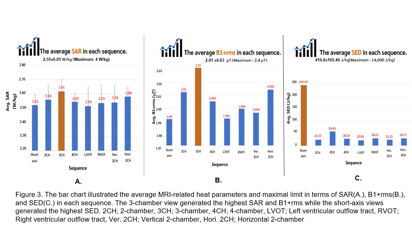
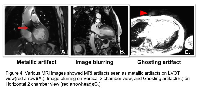
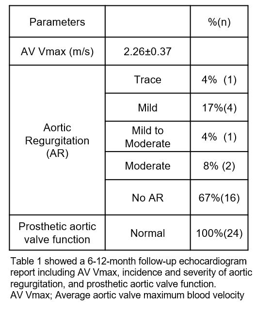
All average MRI-related heating parameters are slightly low to the normal acceptable range. Average SAR is 2.55 ± 0.05 W/kg (Maximal limit is 4 W/kg), B1+rms is 2.01 ± 0.03 µT (Maximal limit is 2.8 µT) and SED is 416.8±165.4 J/kg (Maximal limit is 14,000 J/kg) (Figure 3.). The 3-chamber view and a short-axis view tend to generate the highest heating parameters Factors that affect MRI-induced heating include scan time per slice and total scan time. BMI and body weight were not associated with the heating effect in our study. Good image quality which all cases were able to calculate LVEF. MRI artifacts were found including metallic artifacts (100%), image blurring(75%) and ghosting artifacts(33.3%) (Figure 4.). A 6-12-month follow-up echocardiogram showed normal prostatic valve function in all cases with normal-range AV Vmax. 67% of the patients has no aortic regurgitation while 7% of the patient developed moderate AR (Table 1.) which are proved to be a TAVR-related complication.


Case Summary
Our study is the first frontier pilot challenging study that demonstrates the feasibility, safety, and clinical efficacy of the early 3-Tesla cardiac magnetic resonance imaging within 14 days post-TAVR procedure. The cardiac MR imaging could be carefully added to routine imaging workflow with a safe, good-quality image and provide more information and reproducible structural and functional cardiac evaluation with a good assessment of LVEF despite metallic artifacts. From our standard cardiac MRI protocol and best practice, MRI-related heat parameters are slightly low to the normal acceptable range according to MRI safety guidelines and International Electrotechnical Commission (IEC). Factors that affect MRI-induced heating include scan time per slice and total scan time. BMI is not associated with the heating effect in our study. The 3-chamber view and a short-axis view tend to generate the highest heating parameters. Optimization between image quality, image information requirement, and MRI-related heating effect should be scrutinized and properly tailor-made adjusted depending on the patient's condition and clinical question. No serious MRI-related complications were found either during performed cardiac MRI post TAVR from 3 to 13 days or 6-12-month follow-up from the echocardiogram report. However, a larger sample size, multicentre implementation, and ongoing long-term follow-up should be further investigated for more information.
